
Cubital Tunnel
Syndrome
Jacksonville, FL
Numbness or tingling in the small finger and the ulnar side of the ring finger — especially at night or when the elbow is bent — is characteristic of cubital tunnel syndrome. Jacksonville hand and upper-extremity surgeon R. David Graham, MD treats ulnar nerve compression at the elbow with nonsurgical care, in-situ decompression, and anterior transposition when appropriate.
- Numbness or tingling in the ring and small fingers
- Symptoms worse when the elbow is bent — sleeping, driving, talking on the phone
- Weak grip or difficulty with fine motor tasks
- Pain or aching at the medial (inner) elbow
- Difficulty spreading fingers apart
- Wasting of the hand's intrinsic muscles in advanced cases
Understanding the Condition
What Is Cubital Tunnel Syndrome?
The Nerve Behind Your Funny Bone
Cubital tunnel syndrome is compression of the ulnar nerve at the elbow — the second most common nerve compression syndrome in the upper extremity after carpal tunnel syndrome. The ulnar nerve passes behind the medial epicondyle (the bony bump on the inside of the elbow, often called the "funny bone") through a fibrous tunnel called the cubital tunnel. When this tunnel is too tight, or when the nerve is repeatedly stretched by sustained elbow flexion, the nerve is damaged.
The ulnar nerve controls sensation in the ring and small fingers and the inner border of the hand, and provides motor function to most of the intrinsic muscles of the hand — the small muscles between the metacarpals that allow precise finger positioning, grip, and fine pinch. When the nerve is compressed, both sensory and motor function suffer.
The classic complaint is numbness and tingling in the ring and small fingers, often waking patients from sleep when the elbow is bent. Some patients notice weakness in the hand — difficulty opening jars, dropping objects, or losing fine motor control. In advanced cases, the intrinsic muscles visibly waste and the hand develops a characteristic "claw" posture in the ring and small fingers.
What makes cubital tunnel syndrome distinctively urgent is the biology of the ulnar nerve itself. It recovers more slowly and less completely than the median or radial nerve after prolonged compression. Acting early — before significant muscle weakness or wasting — substantially improves outcomes. Waiting for symptoms to become severe before seeking evaluation is one of the most common and consequential mistakes in the management of this condition.
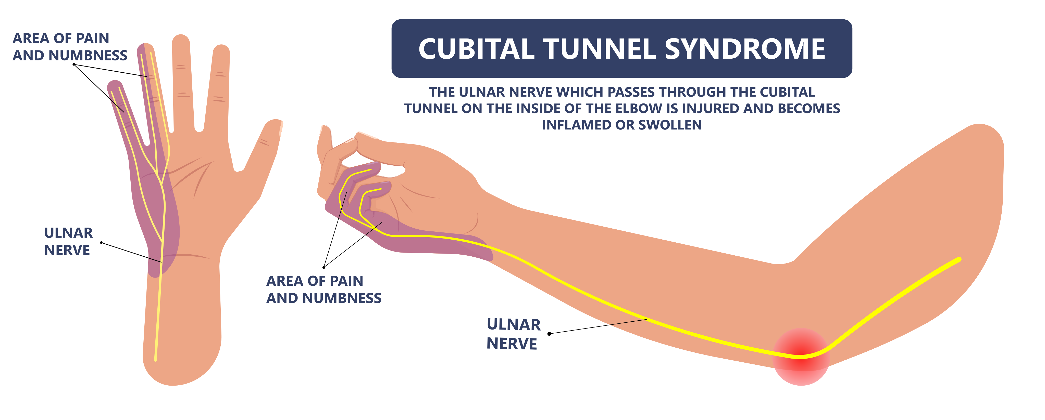
The ulnar nerve passes directly behind the medial epicondyle through the cubital tunnel. When the elbow is bent, the tunnel narrows and the nerve is stretched simultaneously — this is why symptoms are classically worst with sustained elbow flexion: sleeping with the arm bent, talking on the phone, or resting the elbow on a hard surface.
Surgical decompression releases the tight structures compressing the nerve. In-situ decompression leaves the nerve in its natural path. Transposition moves the nerve to the front of the elbow where it is no longer subject to stretch or compression.
When Cubital Tunnel Syndrome
Becomes More Serious
The ulnar nerve doesn't wait well.
Not all nerve compression syndromes carry the same urgency. Carpal tunnel syndrome — median nerve compression at the wrist — can often be observed and treated conservatively for extended periods with good eventual outcomes after surgery. The ulnar nerve is different.
In Dr. Graham's experience, the ulnar nerve is particularly unforgiving once motor weakness develops. Numbness often improves after decompression, but established intrinsic weakness and muscle wasting are less predictable. That is why he prefers to discuss treatment before significant motor loss appears rather than waiting for weakness or atrophy. Once intrinsic muscle weakness develops — once you notice you can no longer spread your fingers easily, or your grip has weakened, or the small muscles between your knuckles have started to flatten — the damage is occurring faster than it can be reversed. Motor recovery after cubital tunnel release is less complete and less predictable than sensory recovery, and it worsens with duration of compression.
Dr. Graham is direct about what the stakes are at each stage. The aim is to make the decision with a clear picture of the findings and the trade-offs, rather than on urgency alone.
With a mild nerve conduction study, the conversation is about prevention. Even mild compression can cause numbness and tingling that becomes permanent if the nerve is not decompressed in time. A mildly abnormal nerve study does not automatically mean surgery is necessary. For patients with intermittent symptoms and no objective weakness, activity modification and nighttime positioning remain reasonable. Dr. Graham also discusses decompression as an option, because he has seen apparently mild disease progress and because established motor deficits can be difficult to reverse. The decision depends on symptoms, duration, examination, electrodiagnostic findings, ultrasound when appropriate, and the patient's preferences. That said, for patients with mild disease who prefer to first try conservative management, Dr. Graham has no objection — conservative measures are entirely reasonable in this group and are always discussed.
With a moderate or severe nerve conduction study, the conversation changes. At this stage, Dr. Graham is direct with patients: the numbness, tingling, and weakness they are experiencing may already be permanent — even with successful surgery. Decompression relieves the ongoing compression and gives the nerve the opportunity to recover, but it cannot guarantee full reversal of symptoms that have been present for months or years. Some patients regain significant function; others stabilize without full return. This is an honest, important conversation that patients deserve to have before surgery, not after.
If you have been told your nerve conduction study shows cubital tunnel syndrome and you are waiting to see if it gets worse — that is the conversation Dr. Graham is prepared to have at your first visit.
Recognizing Cubital Tunnel Syndrome
Cubital Tunnel Syndrome Symptoms
Symptoms follow the ulnar nerve's sensory and motor territory — the ring and small fingers, the hand's intrinsic muscles. They progress from sensory to motor over time.
Numbness in Ring & Small Fingers
The most common presenting symptom. Tingling or numbness in the ring and small fingers — sometimes extending along the inner border of the hand and forearm. Characteristically worse when the elbow is bent and often wakes patients from sleep.
Symptoms with Elbow Bent
Sustained elbow flexion — talking on the phone, sleeping with the elbow bent, driving with the arm resting on a window — reliably triggers or worsens symptoms. This positional component is a hallmark of cubital tunnel and distinguishes it from other nerve conditions.
Inner Elbow Pain or Tenderness
Aching or tenderness directly over the medial epicondyle and cubital tunnel. Tapping over the nerve (Tinel's sign) may produce tingling that shoots into the ring and small fingers. Some patients describe a persistent aching on the inner side of the elbow that worsens with activity.
Weak Grip & Hand Clumsiness
As motor fibers are affected, grip strength weakens and fine motor control deteriorates. Patients notice difficulty with buttons, keys, and precise tasks. This is an important warning sign — motor involvement means the nerve is under significant stress and intervention should not be delayed.
Difficulty Spreading Fingers
The intrinsic hand muscles that allow finger abduction and adduction — spreading the fingers apart and bringing them together — are ulnar nerve-innervated. Weakness in these muscles is an early motor sign. A simple test: can you hold a piece of paper between two straight fingers without bending them?
Muscle Wasting — Advanced Sign
In advanced cubital tunnel syndrome, the interosseous muscles between the metacarpals visibly flatten. The hand looks bony and the spaces between the knuckles hollow out. This is a late finding indicating prolonged, severe compression — and the most important reason not to wait. Wasting is difficult to reverse even after successful decompression.
Diagnosis & Severity
Diagnosing Cubital Tunnel Syndrome
Examination, EMG/NCS & Ultrasound
Three things work together, and none of them settles the question on its own.
Clinical examination. Where the numbness falls, whether holding the elbow bent reproduces it, Tinel's sign at the medial elbow, the strength of the small muscles in the hand, the ability to spread and close the fingers against resistance, and any visible wasting between the thumb and index finger.
Nerve conduction studies and EMG. An NCS measures how quickly the signal travels through the ulnar nerve across the elbow; slowing across the elbow supports the diagnosis and helps quantify severity. EMG examines the muscles the nerve supplies, identifying denervation that indicates motor involvement, and helps distinguish cubital tunnel syndrome from a cervical radiculopathy that can mimic it. A positive study is strong confirmation. A normal study does not exclude cubital tunnel syndrome — early or intermittent compression can produce classic symptoms with normal electrodiagnostics.
Diagnostic ultrasound. Ultrasound shows the ulnar nerve directly. It demonstrates enlargement of the nerve at the point of compression, and because it is dynamic it shows what the nerve does as the elbow bends — including whether it subluxates over the medial epicondyle. That finding is not visible on a nerve conduction study, and it changes the operation: a nerve that dislocates with flexion is the one that needs transposition rather than in-situ decompression. When nerve stability is the question that matters, Dr. Graham obtains a diagnostic ultrasound.
When symptoms persist despite nonsurgical treatment, or when there is muscle wasting, release can be appropriate — the decision rests on those findings combined with the physical examination.
Imaging — X-rays or MRI — is occasionally useful to identify bony spurs, heterotopic ossification, or masses that may be compressing the nerve, but is not required for most cases.
Scroll to see the full table
| Severity | Findings | Dr. Graham's Approach |
|---|---|---|
| Mild | Intermittent numbness, no objective weakness, mild electrodiagnostic changes if present. | Nonsurgical treatment is reasonable. Decompression is also discussed when symptoms are persistent or progressive. In Dr. Graham's experience, apparently mild disease does not always stay mild, and established motor deficits are difficult to reverse. |
| Moderate | More frequent or constant sensory symptoms, early weakness or evidence of motor involvement. | Surgery generally recommended, with counseling that recovery becomes less predictable as motor dysfunction develops. |
| Severe | Constant numbness, significant weakness, intrinsic wasting, clawing, significant axonal loss. | Prompt decompression recommended. Full recovery cannot be guaranteed. |
Non-Surgical Treatment
Nonsurgical Treatment for Cubital Tunnel Syndrome
Nonsurgical treatment is often the first step, particularly for intermittent symptoms. The bar for surgery is appropriately lower for cubital tunnel than for most other conditions — when there is constant numbness, muscle wasting, or nerve damage on testing, waiting is not in the patient's interest.
Nighttime Elbow Positioning
Avoiding prolonged deep elbow flexion at night is a common first-line strategy for intermittent cubital tunnel symptoms. A towel, pillow or elbow splint can keep the elbow from remaining tightly bent during sleep. Dr. Graham commonly combines nighttime positioning with avoiding prolonged pressure on the medial elbow.
Activity Modification
Avoiding sustained elbow flexion — resting the elbow on hard surfaces, cradling a phone with the elbow bent, sleeping with the arm tucked under — reduces repetitive stretch on the nerve. Ergonomic modifications at work (raising a desk, using a headset instead of cradling a phone, positioning the arm on a padded surface) can significantly reduce daily nerve loading.
Elbow Padding
A soft foam pad worn over the medial elbow protects the nerve from direct compression when resting on hard surfaces. This is a simple, inexpensive adjunct that reduces the direct mechanical trauma that can worsen symptoms — particularly for patients who work at desks or rest their arms on hard surfaces throughout the day.
For patients with confirmed cubital tunnel syndrome on NCS — even mild — Dr. Graham's approach is to have a serious conversation about surgery. He has no objection to patients with mild disease choosing to try conservative management first, and he supports that choice fully. But he is honest about the risk: the patients who develop permanent numbness and tingling are often those who waited through a mild nerve study expecting improvement that did not come.
Surgical Treatment
Cubital Tunnel Surgery: In-Situ Decompression vs. Transposition
Dr. Graham's preferred technique is in-situ decompression. Transposition is most commonly used for unstable or subluxating nerves and for selected revisions. Dynamic ultrasound can identify an unstable nerve before surgery, which is part of how that decision gets made. The choice is made based on individual anatomy and history — not habit.
In-situ decompression releases the structures that compress the ulnar nerve at the elbow — the fibrous roof of the cubital tunnel, the Osborne ligament, and any other compressive bands — without moving the nerve from its natural anatomical position. The nerve is decompressed where it sits, which requires less dissection than moving it.
Comparative studies show broadly similar outcomes among the commonly used techniques, so the operation is chosen according to nerve stability, anatomy, prior surgery and the individual patient. Recovery is typically faster than with transposition, and the risk of creating new stretch or kinking on the nerve is avoided.
- Nerve remains in natural anatomical position
- Less dissection than moving the nerve
- Nerve is not relocated, so no new stretch or kinking is created
- No post-operative splint and no routine hand therapy
- Excellent outcomes for primary cubital tunnel syndrome
- Dr. Graham's default for primary cases
Anterior subcutaneous transposition moves the ulnar nerve from behind the medial epicondyle to a new position in front of the elbow, underneath the skin. In its new position, the nerve is no longer subject to compression in the cubital tunnel or stretch from elbow flexion. The nerve is rerouted to a location where it has more room and a gentler mechanical environment.
Dr. Graham performs transposition in two specific situations: revision cases where a prior decompression has failed and the nerve needs to be repositioned, and cases where the ulnar nerve is unstable — meaning it subluxes (snaps over) the medial epicondyle during elbow flexion and extension, causing repeated mechanical trauma.
- Nerve relocated to anterior position — no longer in cubital tunnel
- Eliminates stretch from elbow flexion
- Preferred for revision cases after failed prior surgery
- Indicated when ulnar nerve subluxes over medial epicondyle
- More extensive dissection than in-situ — longer recovery
Anesthesia
Dr. Graham performs cubital tunnel decompression and transposition in the operating room under general anesthesia. Local anesthetic with epinephrine is injected after the procedure is complete — the ulnar nerve sits in a rich venous plexus, and hematoma is a complication worth taking seriously. The epinephrine helps control that bleeding. The procedure takes approximately 30–45 minutes for in-situ decompression.
Incision at the Medial Elbow
A carefully placed incision along the medial (inner) aspect of the elbow exposes the ulnar nerve and the structures compressing it. The medial antebrachial cutaneous nerve — a small sensory nerve that crosses the operative field — is identified and protected. Injury to this nerve is a preventable complication that causes persistent numbness on the inner forearm.
Ulnar Nerve Identification
The ulnar nerve is identified above and below the medial epicondyle and traced through the cubital tunnel. The nerve is assessed for any areas of compression, tethering, or — critically — whether it subluxes over the epicondyle with elbow motion. If subluxation is present, transposition is planned.
Decompression
The Osborne ligament — the fibrous roof of the cubital tunnel — is released. Any additional compressive bands proximally (the arcade of Struthers) and distally (between the two heads of the flexor carpi ulnaris) are also released. The nerve is confirmed to be free of compression through its full course at the elbow and to remain stable in its position with full elbow range of motion.
Closure & Recovery
The incision is closed in layers and a soft dressing applied. The post-operative protocol differs meaningfully between the two procedures:
In-situ decompression: No post-operative splint. The elbow is free to move immediately. No formal hand therapy is needed — patients recover independently, returning to normal activities as comfort allows. Sutures removed at 10–14 days.
Anterior subcutaneous transposition: A post-operative splint is worn for 10–14 days while the wound heals and the transposed nerve settles into its new position. This is followed by a removable brace for an additional 4 weeks. Formal hand therapy is ordered after transposition to restore elbow motion and guide the recovery — the more extensive nature of the dissection makes structured rehabilitation beneficial.
In both cases, nerve recovery begins immediately but proceeds gradually over months — sensory improvement typically precedes motor recovery, and full assessment of outcomes requires 9 months to a year.
Cubital Tunnel vs. Carpal Tunnel
Which Fingers Go Numb?
Both are nerve compression syndromes in the upper extremity, and both cause hand numbness. But they involve different nerves, different locations, different symptoms, and different surgical treatments. They can occur simultaneously — and often do. Understanding which one is causing your symptoms determines the right treatment.
Scroll the table sideways to see all columns
| Feature | Cubital Tunnel Syndrome | Carpal Tunnel Syndrome |
|---|---|---|
| Nerve compressed | Ulnar nerve | Median nerve |
| Location | Behind the medial epicondyle (elbow) | At the wrist — carpal tunnel |
| Fingers affected | Ring and small fingers; inner hand | Thumb, index, middle fingers; sometimes ring |
| Worse with | Elbow bent — sleeping, phone, driving | Wrist position — sleeping, gripping, repetitive use |
| Motor effect | Intrinsic hand muscles — grip, finger spread | Thenar muscles — thumb opposition and pinch |
| Surgical treatment | Cubital tunnel release at elbow | Carpal tunnel release at wrist |
Recovery
What to Expect After Cubital Tunnel Release
Nerve recovery is a biological process that cannot be rushed — but it follows a predictable course. Sensory improvement typically comes first.
Wound Healing
In-situ: No splint — elbow free immediately. No therapy needed. Return to normal activity as comfort allows.
Transposition: Post-operative splint for 10–14 days. Sutures removed at same visit.
Removable Brace Phase
In-situ: Fully active — no further restrictions. Most patients return to all activities within weeks.
Transposition: Splint off, removable brace for 4 more weeks. Hand therapy begins to restore elbow motion and guide recovery.
Nerve Recovery Underway
Both procedures: Sensory symptoms — tingling and numbness — begin to improve. The pace varies by individual and severity of pre-operative compression. Nerve recovery is slow and nonlinear. No activity restrictions for either procedure at this stage.
Long-Term Nerve Recovery — 9–12 Months and Beyond
Much of the clinically apparent recovery can be assessed during the first year, but nerve recovery does not have a strict twelve-month deadline. Sensory and strength improvements can continue beyond a year, particularly in patients who began with severe nerve dysfunction. Recovery is slower and less predictable when significant weakness or intrinsic muscle wasting was present before surgery.
“Dr. Graham performed a Cubital Tunnel release in my left elbow, to relieve the numbness I had been having in my left hand. The procedure went very smoothly and Dr. Graham was very communicative about what to expect in my appointments leading up to the surgery as well as with the follow up the evening after my surgery.”
Common Questions
Frequently Asked Questions
References & Further Reading
The sources below informed this page and are good places to read further. Links open in a new tab.
- Graf A, Ahmed AS, Roundy R, et al. Modern Treatment of Cubital Tunnel Syndrome: Evidence and Controversy. J Hand Surg Glob Online. 2023;5(4):547–560.
- Abourisha E, Srinivasan AS, Barakat A, et al. Surgical Management of Cubital Tunnel Syndrome: A Systematic Review and Meta-analysis of Randomised Trials. J Orthop. 2024;53:41–48.
- Xirou S, Anagnostou E. Electrodiagnosis and Ultrasound Imaging for Ulnar Nerve Entrapment at the Elbow: A Review. Neurodiagn J. 2024;64(4):175–192.
- Florczynski MM, Kong L, Burns PB, et al. Electrodiagnostic Predictors of Outcomes After In Situ Decompression of the Ulnar Nerve. J Hand Surg Am. 2023;48(1):28–36.
- Giladi AM, Gaston RG, Haase SC, et al. Trend of Recovery After Simple Decompression for Treatment of Ulnar Neuropathy at the Elbow. Plast Reconstr Surg. 2013;131(4):563e–573e.
- Palmer BA, Hughes TB. Cubital Tunnel Syndrome. J Hand Surg Am. 2010;35(1):153–163.